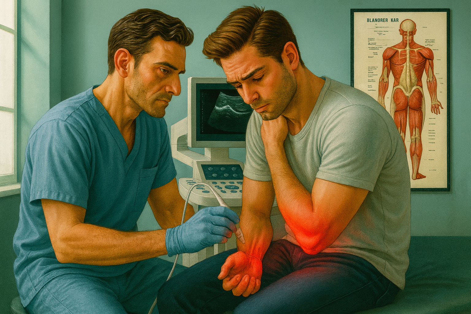
Ultrasound of the musculoskeletal system can contribute to or establish the diagnosis of painful syndromes and allow the accurate, safe and effective application of minimally invasive treatment techniques.
Its application is characteristic for common hand syndromes that afflict active individuals for long periods of time, often leading them to surgery.

Carpal Tunnel Syndrome (CTS) is caused by compression of the median nerve at the wrist level due to anatomical abnormalities (thickened ligament-fascia), systemic conditions (diabetes, pregnancy, etc.), and space-occupying lesions (tendonitis of the flexor tendons, ganglia, etc.).
Symptoms include numbness and burning pain in the thumb, index and middle fingers, and the radial surface of the ring finger. Muscle weakness and atrophy of the thenar muscles (radial surface of the palm) may coexist if the condition has been present for a long time.
The diagnosis can be made based on symptoms, clinical examination, and ultrasound findings. Ultrasound findings may include the area of compression, central swelling of the nerve (cross-sectional area > 10mm2) and peripheral atrophy, widening and loss of its fibrous structure, neovascularization, and increased thickness and curvature of the transverse ligament, and safely establish the diagnosis.
Before surgical treatment and in order to avoid it, the syndrome can be treated with ultrasound-guided perineural injections that aim to decompress the nerve and limit its inflammation and swelling.
A similar decompression technique is applied for conservative treatment of trigger finger syndrome where the area of tendon entrapment is seen with ultrasound in dynamic examination during its movement and is decompressed with injection of a solution through a needle that is placed with ultrasound guidance for accuracy and safety.
The syndrome is caused by continuous strain such as holding a tool for a long time but also in the context of rheumatoid or gouty arthritis, diabetes, Dupuytren’s contractures, and is more common in women and at ages 40-60. It presents as pain at the base of the finger with painful ejection-blocking of the finger when trying to bend or extend it.
The above minimally invasive treatment is applied when conservative treatment with reduced activity, splint and non-steroidal anti-inflammatory drugs does not work or in parallel with it for rapid treatment and remission of the problem and avoiding the need for surgical treatment.
In the case of tendinitis of the extensor digitorum brevis and the abductor digitorum longus (De Quervain’s syndrome) the pain is localized on the radial surface of the wrist. Similarly, ultrasound is used for diagnosis and minimally invasive treatment to avoid the need for surgery.